
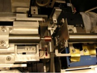
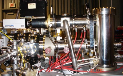
Endstations
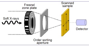
STXM
A Fresnel zone plate is used to focus monochromatic X-rays to a small spot size (~30 nm). The sample is raster scanner through the focal point while detecting transmitted photons. While the technique is mainly bulk-sensitive, since samples tend to be very thin to allow adequate transmission in the soft X-ray region (e.g. ~100 nm for C 1s), surface adsorbed species can often be detected.
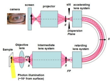
X-ray Photoemission Electron Microscope (X-PEEM)
Photoejected electrons are magnified and imaged with a spatial resolution of ~50 nm. The environment is ultrahigh vacuum, and samples must be conducting and extremely flat. This surface sensitive full field technique features two contrast modes:
X-ray absorption spectroscopy(NEXAFS) - images are generated using the main low kinetic energy peak. Photon energy is scanned. Typically 5-10 nm sampling depth.
Photoelectron spectroscopy (XPS) - images are generated using energy selected electrons. Three possible scan modes:
- scan kinetic energy of electrons with fixed photon energy (XPS)
- scan photon energy with fixed electron kinetic energy (CFS)
- scan photon and electron kinetic energy simultaneously (CIS)
Cryo scanning transmission X-ray microscope (Cryo-STXM)
A cryo scanning transmission X-ray microscope, the cryo-STXM, has been designed and commissioned at the Canadian Light Source synchrotron. The instrument is designed to operate from 100 – 4000 eV (λ = 12.4 – 0.31 nm). Users can insert a previously frozen sample, through a load lock, and rotate it ±70° in the beam to collect tomographic data sets. The sample can be maintained for extended periods at 92 K primarily to suppress radiation damage, and a pressure on the order of 10-9 Torr to suppress sample contamination. The achieved spatial resolution (30 nm) and spectral resolution (0.1 eV) are similar to other current soft X-ray STXMs, as demonstrated by measurements on known samples and test patterns. The data acquisition efficiency is significantly more favorable for both imaging and tomography. 2D images, 3D tomograms and 4D chemical maps of automotive hydrogen fuel cell thin sections are presented to demonstrate current performance and new capabilities, namely cryo-spectrotomography in the soft X-ray region.

(a) Schematic diagram of Cryo-STXM, 1: Zone plate, 2: OSA, 3: Sample, 4: Detector; (b) Conventional mode; (c) Goniometer mode.